Histological Sectioning
Of the three animals collected for this study, one was selected for a radula extraction and another to be fixed for sectioning. Unfortunately the radula extraction was unsuccessful, with the radula not able to be detected. The second animal was fixed at Heron Island for later sectioning and staining at the University of Queensland as described below, with the resultant images of the sections presented here.
The specimen was first relaxed in Magnesium chloride to ensure that the parapodia were outstretched, then cooled in the freezer for ten minutes before being placed in 4% paraformaldehyde (PFA) in seawater, and then into a refrigerator overnight. The animal was then stepped up into 75% ethanol over a half hour. 25% of the total volume was removed and replaced with ethanol. This was then swirled to mix the contents, and left to stand for five minutes. The process was then repeated removing 50% of the total volume followed by 75%, then finally 100%. Once fixed, the animal was left in ethanol for transport from Heron Island to Brisbane for sectioning. The animal was mounted in wax and transversely sectioned in an anterior direction, beginning at the posterior aspect of the animal. The sectioned slides were then stained with haemtoxylin and eosin (H & E) and viewed microscopically. The images obtained from the sectioning of Elysia sp. are discussed below.
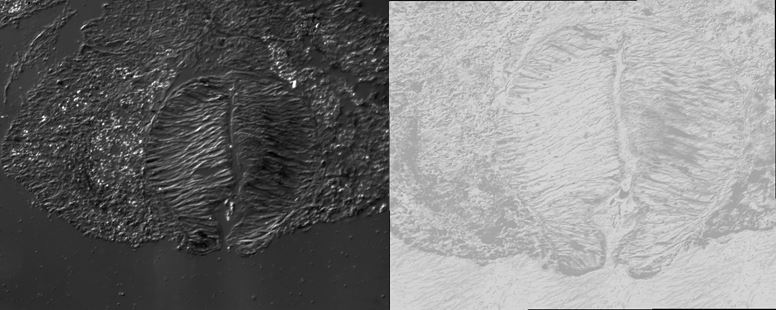
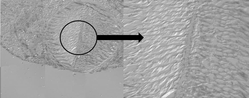
Figure 1 shows a cross section of Elysia sp. taken from the posterior end of the animal, with the parapodia outstretched laterally, and the body in the middle. Although not entirely clear in this image, the greatest nuclei densities are seen just above the foot and within the parapodia. Figures 2 and 3 below show greater detail of the foot, which is highly muscularised in Elysia sp. Figure 2 illustrates a section in which the foot has curled up slightly, with the transverse groove quite prominent. The section seen in Figure 3 illustrates an uncurled foot, and on the right hand side an image of the transverse groove itself as viewed under 40x magnification.
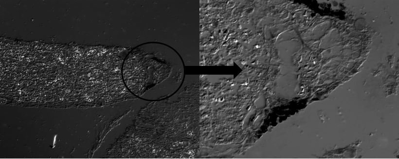
Upon viewing the microscope slides, black pigmentation was noted on the innermost edges of the parapodia on some of the sections as seen in Figure 4 below. Initially it was thought that this was colour pigmentation from the slug, however upon comparing the sections to the morphology of the slug, this does not seem to be in agreeance with the location of the black pigmentation seen on Elysia sp. In some Elysiids, digestive gland tubules extend laterally into the parapodia, even in the posterior end, while in others hermaphroditic follicles are thought to extend into the parapodia also, though is it likely that this is more commonplace in the anterior end of the animals [1]. Given the close proximity to the body, it is possible that the pigmentation seen in some of the sections could be an extension of the digestive gland tubules, though further analysis with more specimens would be required to confirm this.
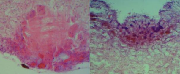
It was clear from viewing the slides that the highest nuclei densities were seen in the upper epithelial layers of the dorsal surface of the body, and surrounding the foot. This is best illustrated in Figure 5 below, where the darker purple regions are the nuclei.

Figure 1- Stitched image of a cross section of Elysia sp. from posterior end of animal. The nuclei are seen to most dense above the foot and in the parapodia.

Figure 2- In these sections the foot has curled up slightly and the transverse groove is quite prominent, as viewed at 5x magnification.

Figure 3- The foot of Elysia sp. is split by a tranverse groove, as seen on the left (stitched image), and as viewed at 40x magnification on the right.

Figure 4- Cross section of Elysia sp. showing pigmentation on the parapodia at 10x (left) and 40x (right) magnification.

Figure 5- Cross section of Elysia sp. at 10x magnification demonstrating high densities of nuclei (purple) surrounding the foot (left) in the epithelial layer of the dorsal surface of the body (right).
|